
One shoulder slightly higher, a protruding shoulder blade, an asymmetric waist noticed by the paediatrician or in the mirror. Or, in adulthood, back pain that will not go away and a leg that hurts when walking. Behind such different situations there may be the same word: scoliosis.
On this page I answer the most frequently asked questions, distinguishing between the two main situations: adolescent scoliosis, which is discovered during growth, and adult scoliosis, which often presents with pain. At the end I explain, step by step, how the operation is performed when it is needed.
What is scoliosis?
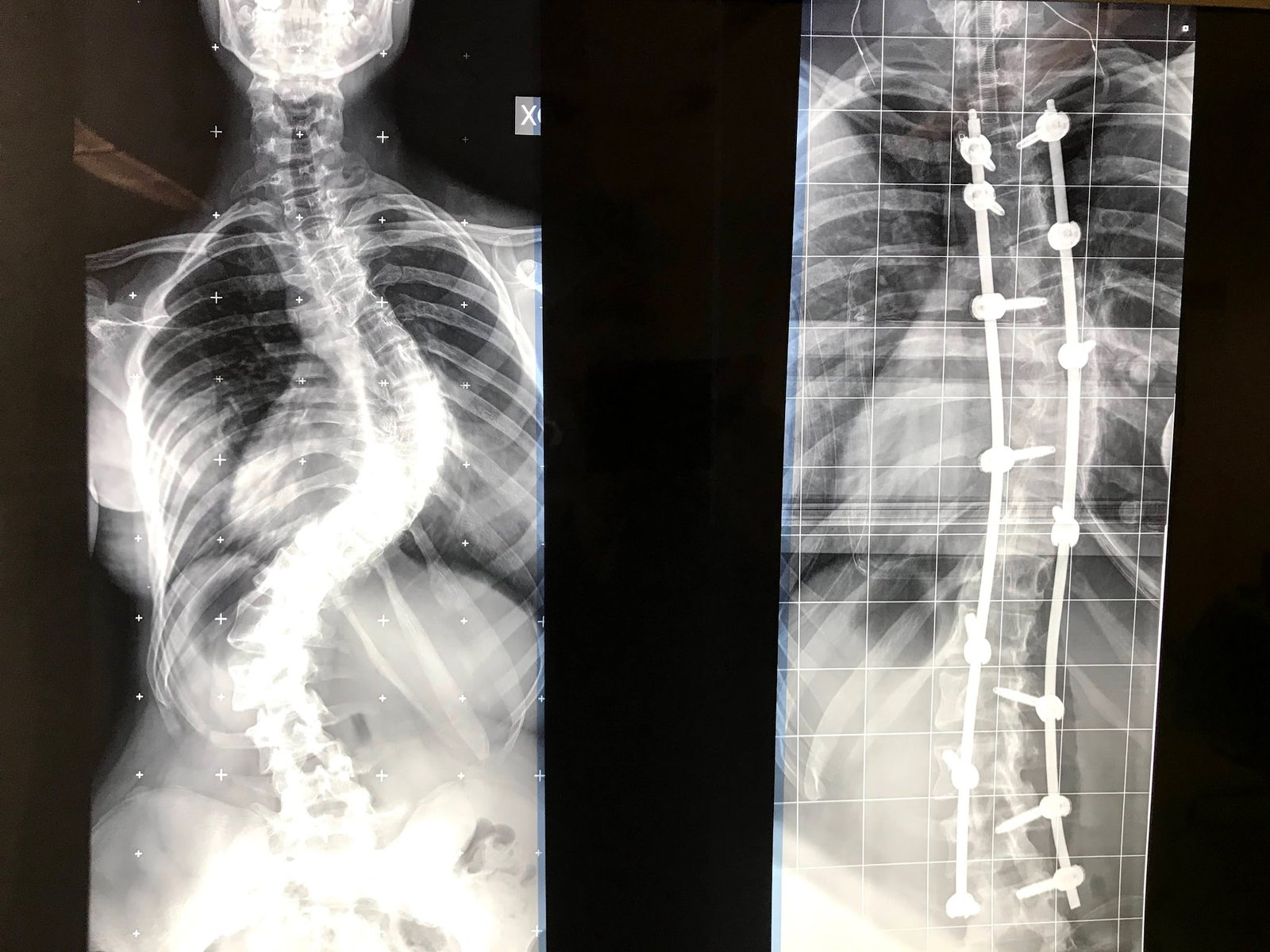
It is a three-dimensional deformity of the spine: not just a sideways curve seen from behind, but also a rotation of the vertebrae, which is why it is often called rotoscoliosis. The rotation is what creates the hump, the prominence of the ribs or lumbar muscles when bending forward. Scoliosis is diagnosed when the curve exceeds 10 degrees, measured using theCobb angle on the X-ray.
Adolescent scoliosis
Why does it affect young people?
In most cases it is idiopathic scoliosis, i.e. without a specific cause: genetic predisposition plays a role, and it appears or worsens during the pubertal growth spurt. It is more common in girls, who also have a higher risk of curve progression (Weinstein et al., Lancet 2008). Backpacks, posture at the desk or “asymmetric” sports do not cause scoliosis.
How is it detected?
With a simple test: the Adams forward bend test. The young person bends forward with arms hanging down and the back is observed from behind: a hump on one side is the signal for further investigation. Confirmation comes from a full-length standing X-ray of the spine, on which the Cobb angle is measured and the remaining skeletal growth is assessed (Risser sign).
Does bracing really work?
Yes. The BrAIST study (Weinstein et al., N Engl J Med 2013) showed that, in young people who are still growing and have moderate curves, bracing significantly reduces progression to the surgical threshold, and that the benefit increases with the number of hours per day the brace is worn. The SOSORT guidelines (Negrini et al., 2018) recommend a stepwise approach: observation, scoliosis-specific exercises and bracing according to the size of the curve and the remaining growth.
When is surgery needed in adolescents?
When the curve exceeds roughly 45–50 degrees, or continues to worsen despite bracing. Curves of this size tend to progress even after growth has finished and, in the most severe cases, can interfere with breathing. The aim of surgery is to correct and halt the curve with spinal fusion (arthrodesis).
Adult scoliosis
What is adult scoliosis?
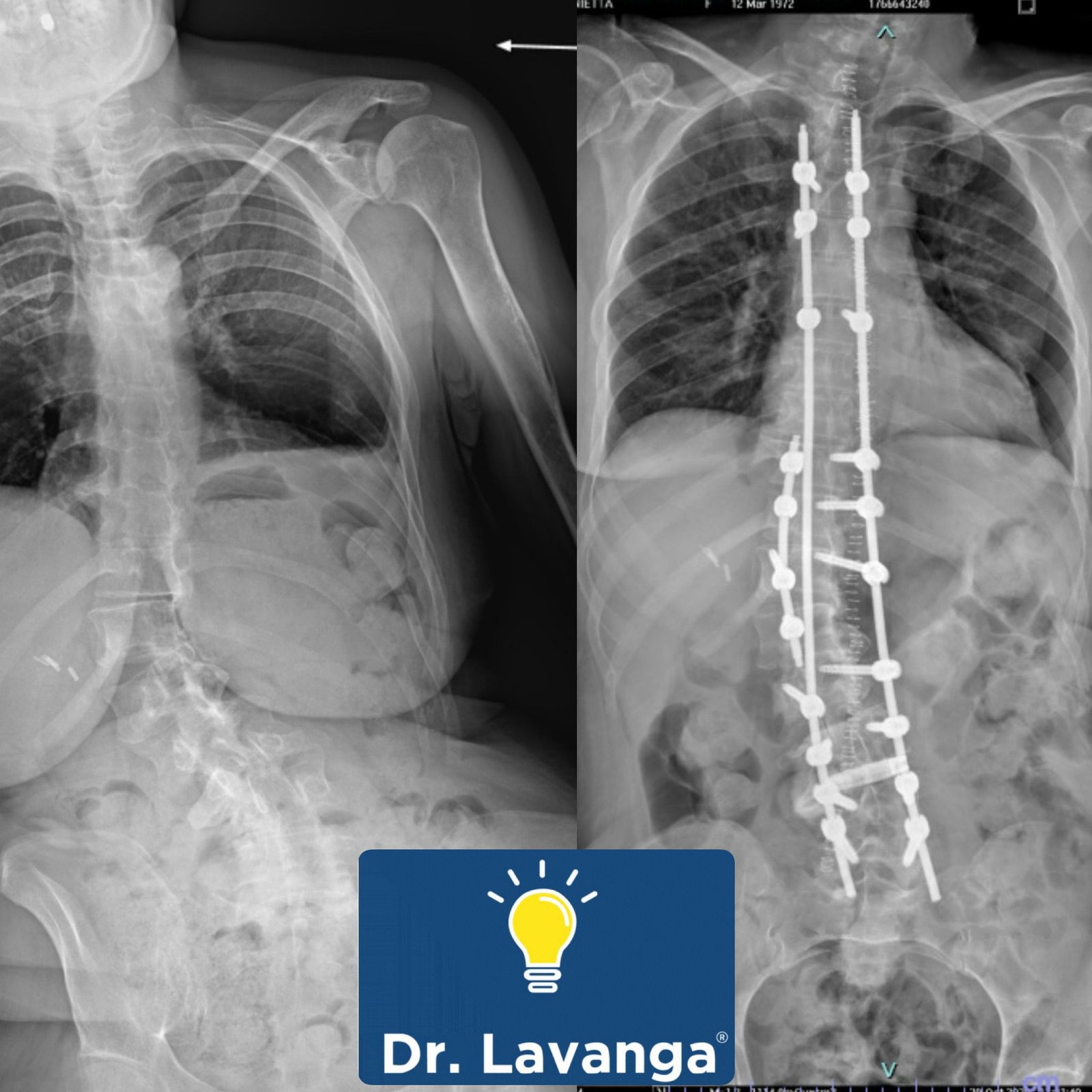
Aebi (Eur Spine J 2005) classifies it into three main groups: “de novo” degenerativescoliosis, which develops after the age of 50 because of asymmetric wear of the discs and joints; adolescent idiopathic scoliosis that progresses into adulthood; and secondaryforms, for example due to osteoporosis, previous surgery or differences in leg length.
Why does it hurt and cause sciatica in adults?
Because the curve is accompanied by disc degeneration, facet joint arthritis and, often, stenosis of the canal or foramina on the concave side of the curve. The result is back pain, leg pain when walking, difficulty standing upright and, in forms with loss of lordosis, the feeling of “falling forward”. See also the page on assessing back pain.
Surgery or conservative treatment: what do the studies say?
The prospective multicentre study by Bridwell (Spine 2009) followed adults with symptomatic lumbar scoliosis for two years: those who had surgery showed a significant improvement in pain and quality of life, whereas those who received conservative treatment alone did not, on average, improve. This does not mean that everyone should have surgery: physiotherapy, exercise, medication and injections remain the first step, but when pain is disabling, surgery has a documented role.
What is the role of minimally invasive techniques?
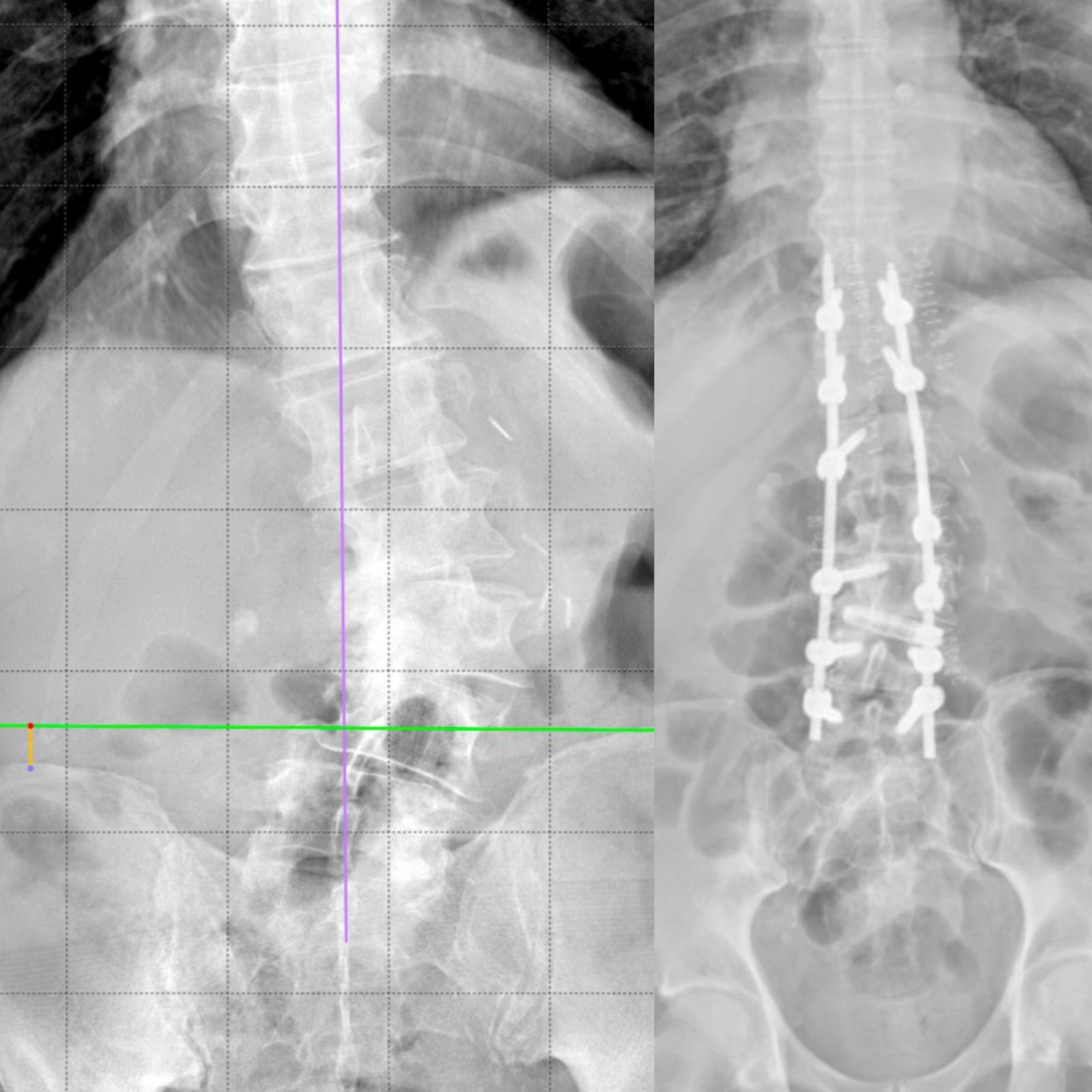
In adults, whenever possible, I combine posterior correction with interbody cages inserted via a lateral approach (XLIF), which reopen the disc spaces, indirectly decompress the nerves and correct the curve with less bleeding. Planning is always personalised: age, bone quality, sagittal balance.
How the operation is performed, step by step
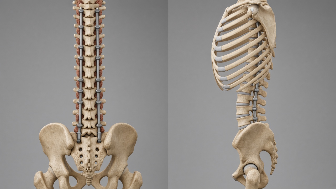
When surgery is indicated, the most commonly used technique isposterior spinal fusion (arthrodesis) with pedicle screws and rods. Here are the main stages, simplified.
1. Planning and positioning
I study the curve on full-length standing and side-bending X-rays and on CT/MRI, and decide which levels to include. In the operating theatre the patient lies face down, with continuous neuromonitoring of the spinal cord and nerves throughout the operation.
2. Insertion of the pedicle screws
Through an incision along the midline of the back, the screws are inserted from back to front, through the pedicle, into the vertebral body. The trajectory is guided by neuronavigation (O-arm) for maximum precision, away from the spinal cord and nerve roots.
3. Rods and curve correction
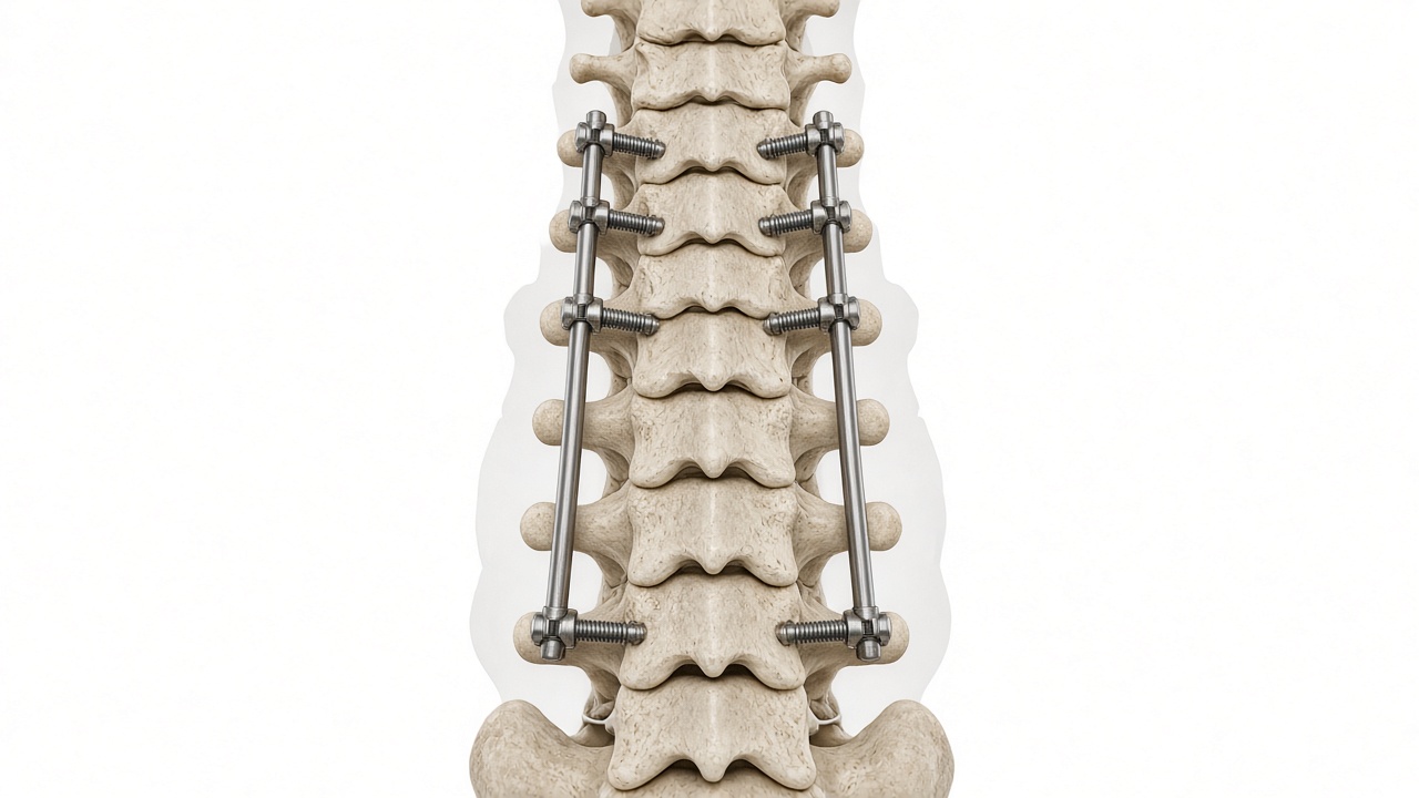
Two rods made of titanium or cobalt-chrome, shaped to the corrected profile of the spine, are attached to the screw heads. Using rotation, translation and compression/distraction manoeuvres, the curve is reduced and the natural curves in the side view are restored.
4. Bone fusion and closure
The posterior bone surfaces are prepared and enriched with bone graft: over the following months the vertebrae fuse together into a single solid block —spinal fusion (arthrodesis). The screws and rods hold the correction until the bone has consolidated. In adults, interbody cages are added at some lumbar levels, including via a lateral approach (XLIF).
5. After surgery
Patients usually get up within the first few days with the physiotherapist; discharge usually follows after a few days. School or office work is generally resumed after 4–8 weeks, and sport gradually after a few months, depending on X-ray check-ups.
Other frequently asked questions
Which tests are needed?
Full-length standing X-rays of the spine (front and side views), side-bending X-rays if surgery is being considered, an MRI if there is leg pain or neurological signs, and a DXA bone density scan in adults to assess bone quality.
Can people with scoliosis play sport?
Yes; in fact, sport is recommended. No sport “causes” or “cures” scoliosis on its own; specific exercises are part of conservative treatment.
Pregnancy and scoliosis?
Scoliosis, even after surgery, does not usually prevent pregnancy or natural childbirth. It should be mentioned to the anaesthetist if an epidural is being considered.
Does scoliosis get worse with age?
It depends on the size of the curve. Small curves at the end of growth usually remain stable; curves over 40–50 degrees can continue to worsen slowly in adulthood too, by about one degree per year. In adults, degeneration of the discs and joints can also cause curves that develop after the age of 50 to progress. This is why we recommend regular X-ray check-ups for significant curves.
Does the back stay stiff after spinal fusion (arthrodesis)?
The fused segment no longer moves, but the rest of the spine, the hips and the shoulders compensate. Many patients return to an active life, including sport. The goal is to choose the minimum number of levels needed to achieve a balanced, long-lasting spine.
Like a mountain ridge
A ridge is never straight, and it does not need to be. The spine also has its natural curves: our job is not to make it rigid, but to restore its balance.
Book a consultation
Have you noticed an asymmetry in your child, or do you suffer from back pain with known scoliosis? Write to me on WhatsApp at 392 39 91 841 or contact the Ortho-Next secretary's office: segreteria@orthonext.it, tel. +39 331 185 9007. Free guide to back exercises: Write GUIDA on WhatsApp.
Educational content: this does not replace a medical consultation, diagnosis or personalised treatment.
References
- Weinstein SL et al. Adolescent idiopathic scoliosis. Lancet. 2008. PMID 18456103 · DOI 10.1016/S0140-6736(08)60658-3
- Weinstein SL et al. Effects of bracing in adolescents with idiopathic scoliosis (BrAIST). N Engl J Med. 2013. PMID 24047455 · DOI 10.1056/NEJMoa1307337
- Negrini S et al. 2016 SOSORT guidelines: orthopaedic and rehabilitation treatment of idiopathic scoliosis during growth. Scoliosis Spinal Disord. 2018. PMID 29435499 · DOI 10.1186/s13013-017-0145-8
- Aebi M. The adult scoliosis. Eur Spine J. 2005. PMID 16328223 · DOI 10.1007/s00586-005-1053-9
- Bridwell KH et al. Does treatment (nonoperative and operative) improve the two-year quality of life in patients with adult symptomatic lumbar scoliosis: a prospective multicenter evidence-based medicine study. Spine. 2009. PMID 19752703 · DOI 10.1097/BRS.0b013e3181a8fdc8